acetabulum anatomy

Acetabulum Of The pelvis
One of the most common injuries that a person in a wheelchair leads to is a fracture of the acetabular region.Today we learn that this is associated with the hippocampus, as well as some treatments for dysplasia or other problems there. Find out also where problems with acetabulum can lead to sclerosis or fractures.
WHAT IS HIP? acetabulum anatomy
It is very powerful and the largest in the human body. In addition to flexion and extension features, abduction, forward, side, rotational movements, it also increases backward on the hip flexors.
The unique characteristics of this common - they provide approximately 40% of human activity.
This acetabulum is also building up in our head, as well as depression. Dale is as deep as their common shoulder.Both cover its cartilaginous tissue, which can absorb the load, smooth movement while skipping its member, running, and T. D.
THE STUDY OF BODY PARTS
Acetabulum is a break in the iliac bone, part of the hipbone. It performs some of the most important and complex tasks in the body as support and movement. It has a hemispherical shape that is left on the inner cartilage. Dorsal wall of the front and acetabulum of the doctors, and the arch isolated. The pelvis is given a human activity in this section, which is important to find and treat injuries quickly in this area.
Acetabulum is made up of the bones of the chest, ischium, and ilium in place of their connection.
acetabulum anatomy
FRACTURE
Too often, bone continuity occurs as a result of such a violation. In addition, this injury can occur after a great height.
There are 2 types of fractures in the acetabulum:
- Light damage. This is caused by the front columns, the posterior and middle wall transverse damage.
- Complex damage. Only a few fragments of bone on the line they reach are at this time of amazing. This includes front wall damage, transverse, columns and both.
Symptoms of a fracture include:
- Pain in the brush and hips.
- It is difficult for the patient to rely on the injured foot.
- For athletes, which is cutting off the hips and knees, a clear advantage for shortening the. The leg outwards turned.
acetabulum anatomy
TREATMENT OF FRACTURES
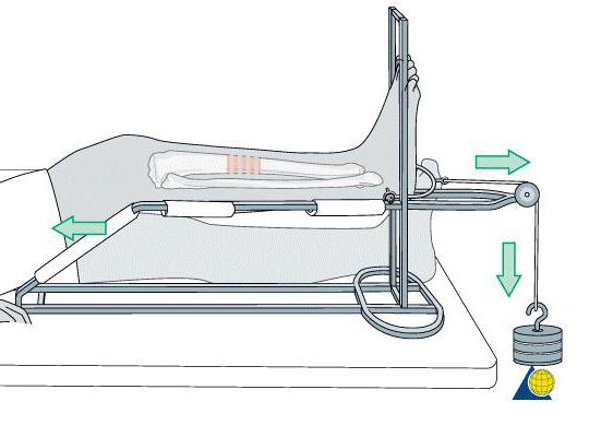
- If a violation of the spine's position occurred without discrimination, then the patient underwent a special bus, a tape and a special extension of the lower leg for about 1 month. Be sure to appoint Electrophoresis Physiotherapy Pathway.
- If the upper and posterior margins of the acetabulum of the pelvis are broken, then treatment is restored to the bone marrow, hip replacement.The epicondyles that lead to a specialist speak for you. The common face is that this accession is extended thanks, and when it is pressed on the remaining acetabulum, the adhesive is present. It is usually 1.5 months when you extend your stay.
acetabulum anatomy
If a fracture between a large and impossible to compare, surgery is necessary. This is not to be done within the first two weeks after injury. To fix the pool crater, use a surgical plate and distressed screws.
After treatment, it is very important during the bone regeneration.
POSSIBLE ACCESS METHODS
Acetabulum fractures, such as relaxation due to relaxation, are very challenging. This is very difficult to say to cause a specialist injury.
Of course there are many types of fractures involved, and each destination has its own method b. The following methods are used primarily:
- Front Access.
- ilioinguinal route.
- Back access.
FRONT PATH
In another way it is called the "ofemoralnaya path". All of this fracture is used to rotate to produce an open front wall and an acetabulum column. Additional frontal transverse fractures may be applicable in surgery.
ILIOINGUINAL ACCESS
This is used by opening the front and acetabulum in the inner surface. In addition, single-stage fracture stabilization of the sacroiliac joint can be used to relax and use the eyes.However, this method does not provide people with access to a trained rear control column and wall.
THE BACK ROAD
After mentioning the typical hip displacement, if there is damage to the posterior wall of the acetabulum, it will look for reduction and osteosynthesis. In addition, this method is used to remove joint and cartilage areas.
THERAPY OF THE LATERAL RIB FRACTURE
There is a change if such a disruption is taken during the fall or height of the fall. Young people most people are exposed to this injury. Fractures, dislocations, accompanied by dislocations of the bones of the bones, damage to the cartilage of the articular. Observed in neutral conditions the edge of the acetabulum of Persia. Most posterior column fractures were found.
acetabulum anatomy
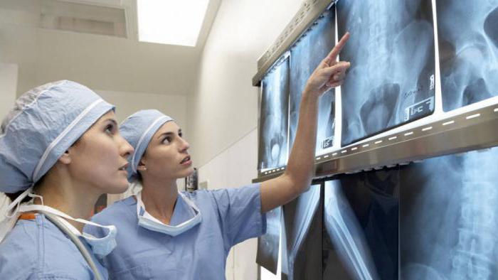
Using a study by Eid X-rays by a hospital specialist, we will investigate the victim. As epidural anesthesia or anesthesia is an urgent issue, there is a reduction in displacement. Later, the final diagnosis of joint damage, including ileum, obstruction review, X-ray and computer tomography,.Such research methods help acetabulum experts to get a complete picture of what they can produce, such as injury.
He does this and just puts someone on foot in surgery. The doctor makes the cut for line where the piece is translated. Then the doctor splits it or loosens it. Stability is in the process of stabilizing fragment stabilization;Then the wound opened.
ACQUISITION
When establishing the acetabulum of the pelvic bone after violating our position, it is important to recover the following rules:
- Daily discount with special breathing exercises.
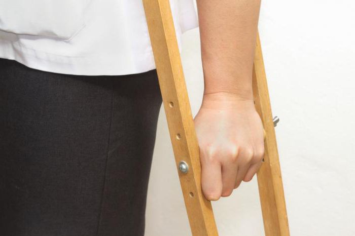
- Learn the right to walk on crutches, provided that they are on foot.
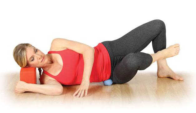
- Provide a unique collection of activity under the supervision of a podiatrist: flexion and toes, extending the pelvis to support a healthy bend to the foot, between the toes and the two hands.
THEY'RE IN TOUCHDOWN
The only symptom of the disease is acetabulum, multiple sclerosis, seen in X-rays. This term is often found in X-ray images of product images.
This problem develops due to inflammatory changes in the bones with the overgrowth of the tissue.
Acetabular sclerosis - a condition in which there are no external symptoms - arthrosis. This problem is common in older people. The main causes of depression are sclerosis:
- implants between cartilage.
- Violation of blood supply to the feet with diseases related to the body.
- Arthritis, osteochondrosis are inherited naturally.
- Dislocations in the foot.
- Sit down.
- Joint and congenital anatomy.
- Use injured ligamentous equipment during sports loads injury.
- Fractures in joints.
- Obesity.
DALE, MS TREATMENT OF HERPES
Treatment includes:
- Massage.
- He buys (a hammer in a vault).
- Physiotherapeutic processes (mineral wax, magnetic).
- Special baths with radon, receive hydrogen sulfide.
- Treatment problems NSAIDs "diclofenac", "Nimesulide" and more.
It should also be restricted to lifting the weight to sit for a long time.Jumping, running is forbidden.
OTTO DISEASE
In another way, this disease is called "acetabular dysplasia". And the name of this disease, as the name of the disease, Otto, is a congenital disease that occurs only in women and is found later in 1824, who discovered it later. The problem (in the lower limbs, abduction, adduction, rotation) is manifested in the limits of movement between the joints. In this case, fair sex is no pain.
It is important to conduct a survey to confirm "depression dysplasia":
- Dale in the forecast that requires X-ray in common.
- MRI.
- Ultrasound.
ACETABULUM: OTTO DISEASE TREATMENT
Treatment may include surgical intervention;
- Reduced displacement.
- Rectal surgery for Hiari.
- Open displacement is reduced.
- Bone tract.
- Hip arthroplasty.
Additional treatments are also used:
- A special kind of wrap.
- Therapeutic Practice, Gymnastics.
- Massage.
- Treatment with medicines.
BROKEN WITH PROBLEMS
Motivation in the displacement of the acetabulum, a car accident, for example, or, in the direct plane crash that can occur in the fall of a large object in the pit.
In these complex fractures, the lines between the hip joint were broken.When posterior dislocation performs more trochanter. If he was to be evacuated to the center, he was shot and given a shot. To understand the displaced fracture, it is necessary to do an x-ray in two projections because this problem can be in the front or rearward.
Symptomatology issues:
- Active foot movement is severely restricted.
- It is in a state of low hand and cruelty.
In this case treatment is:
- System of bone contact application.They spoke carrying a supracondylar region in the thigh with a mass of 4 kg.
- The flexion of the foot is placed in a position to bring in the hips and knees.
- Experts to determine the head to perform head-to-head compression by bone graft or bone traction to the desired location of the desired location.
- Reduced loads are transferred to the bone tract leaving the source sinker on the neck axis.
- An angle of 95 degrees is given an angle of 1 week.
It starts at 8 10 weeks when you extend your stay. Even later, 2 weeks are allowed in the joint. Only full load on foot is allowed after six months.And the job skills are in 7 months.
COXARTHROSIS
This disease is characterized by a debilitating nature that affects the elderly and the middle ages. For years, slowly developing disease.
The symptoms of Koksartroza are:
- Pathological relationship between the femoral head and acetabulum.
- The head is on the side of the medial quadrant.
- The roof of the acetabulum imbricate is attached to the cavity, which is visible to the beak.
- Rescue length and the roof is broken.
- thickening of the cortical layer in the roof depressions.
Coxarthrosis pain and limiting joint activity.
In the later stages of the disease there is no thrombosis in the muscles.
The causes of this disease are divided into 2 types;
- Primary coxarthrosis.Which occurs for unknown treatment.
- Secondary coxarthrosis. It is found to be due to other diseases.
It can be the result of problems like the latter type of disease:
- They release hip hop for the man.
- Hip dysplasia.
- Aseptic necrosis of the femoral head.
- Perthes disease.
- Traumatic (broken hip, pelvis, sprains).
Because coxarthrosis was progressing. If you start treatment at an early stage, then you can practice conservative medicine. Only an effective method at a later stage will only have surgery.
TREATMENT OF COXARTHROSIS
Treatment of the disease includes orthopedists. The choice of treatment depends on the stage of the disease.
In Grades 1 and 2 1. The following treatment is assigned:
- Adoption of anti-inflammatory drugs. Do not use them, however, as they can have a negative impact on the internal organs for a long time.
- (use "Arteparon", "rumalon", "chondroitin," "Struktum" as a drug) hondroprotektorov
- vasoconstrictor drugs ("Trental", "Cinnarizine").
- Medicinal muscles relax.
- articular injection, with the use of hormonal agents, such as "kenalog", "hydrocortisone".
- Use perfume additives.
- Physiotherapy (laser-, light therapy, UHF, magnetic therapy), as well as the interval between balsam, specialized gymnastics.
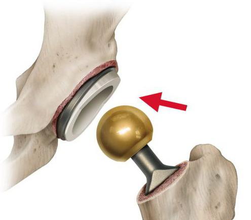
2. The operation is the only way for me to require coxarthrosis in 3rd place. The patient replaces the common endoprosthesis. The repair is performed in one planned manner in general anesthesia. The open patient will be removed on the 10th day after the outpatient treatment.After the repair, recovery steps are necessary. Almost 100% of the injured hip joint provides complete restoration of the function of the injured limb. Even in active activity you will play sports even if you can work people at the same time. If you can wear an artificial body, if you are all 20 years old, the doctor can advise.Repeated operation is necessary to replace a premature endoprosthesis long after this operation.
ACETABULAR LESIONS COMPLEX
Problem: Near the road, it can happen occasionally, but we still need to know about people. Postoperative complications include:
- Exposed.
- Causing wounds.
- Thromboembolism.
- Damage to the nerves.
- aseptic necrosis of the femoral head and acetabulum wall.
- Paralysis of small and medium glute.
To prevent such complications, many doctors immediately provide patients with arthroplasty.
CONCLUSION
During rotation, this acetabulum is characterized by very important early detection, including X-ray, ultrasound, magnetic resonance imaging. Operation - a strict conservative or aggressive: based on these studies; The doctor should choose the appropriate treatment method. Man walks on the complexity of the fast-paced complexion, because it is also important during the course of therapy and recovery.



No comments:
Post a Comment
Welcome to healthcare management